10 Feb Challenging Cases Accelerated Breast Cancer Radiation Therapy February 20, 2023 By drvineeta 0 comments Facebook Twitter WhatsApp WhatsApp Dr Vineeta Goel, Director, Radiation Oncology, Fortis Hospital, Shalimar Bagh Breast cancer treatment has undergone considerable evolu...Continue reading
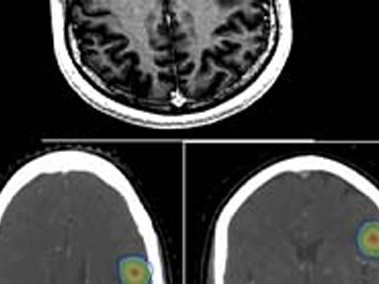
10 Feb Challenging Cases Stereotactic Radiation Therapy For Brain Metastases February 20, 2023 By drvineeta 0 comments Facebook Twitter WhatsApp WhatsApp Case 1 -Single Brain Metastases A 55 Years old lady was diagnosed with left breast cancer in August 2015. She was treated with breast ...Continue reading
10 Feb Challenging Cases Radiation Therapy For (Left) Breast Cancer With Cardiac / Heart Protection February 20, 2023 By drvineeta 0 comments Facebook Twitter WhatsApp WhatsApp Breast Cancer is one of the most common cancers among women. Good news is, that many more patients today are long term breast cancer “s...Continue reading
10 Feb Challenging Cases Breast Cancer Radiation Therapy February 20, 2023 By drvineeta 0 comments Facebook Twitter WhatsApp WhatsApp Radiation Therapy (RT) for breast cancer has seen a long journey precision and focused therapy. RT is an important part of breast cance...Continue reading
10 Feb Challenging Cases Stereotactic Radiotherapy February 23, 2023 By drvineeta 0 comments Facebook Twitter WhatsApp WhatsApp Conventional radiation therapy for cancers often results in some collateral damage to nearby healthy tissues. Stereotactic Radiotherapy...Continue reading
10 Feb Challenging Cases Stage IV Breast Cancer With Paralysis Of Lower Limbs February 20, 2023 By drvineeta 0 comments Facebook Twitter WhatsApp WhatsApp A 48 years old woman presented to us with paralysis of 4 days duration. She also gave a history of upper backache since past 5 months. ...Continue reading
10 Feb Challenging Cases Total Skin Electron Beam Therapy February 20, 2023 By drvineeta 0 comments Facebook Twitter WhatsApp WhatsApp Total Skin Electron Beam Therapy (TSET) is a radiation technique which is used to treat patient’s entire/total skin surface using elect...Continue reading
10 Feb Challenging Cases Solitary Brain Metastases From Breast Cancer-Treatment With Stereotactic Radiation Therapy February 20, 2023 By drvineeta 0 comments Facebook Twitter WhatsApp WhatsApp A 54 Years old lady was diagnosed with left breast cancer in August 2015. Her breast biopsy confirmed it to be Invasive Ductal Carcinom...Continue reading
10 Feb Challenging Cases Intra Operative Radiation Therapy (IORT) With Brachytherapy February 20, 2023 By drvineeta 0 comments Facebook Twitter WhatsApp WhatsApp Intraoperative radiation therapy (IORT) is single dose of focused radiation therapy delivered at the time of surgery. Brachytherapy is...Continue reading
10 Feb Challenging Cases Management Of Intracranial Non Germinomatous Germ Cell Tumour (Choriocarcinoma) Brain With Chemotherapy And Cranio Spinal Radiation Therapy February 20, 2023 By drvineeta 0 comments Facebook Twitter WhatsApp WhatsApp 18 Years old college student presented to hospital with complaints of severe headache, blurring of vision, vomiting and episode of unco...Continue reading