Accelerated Breast Cancer Radiation Therapy
Dr Vineeta Goel, Director, Radiation Oncology, Fortis Hospital, Shalimar Bagh
Breast cancer treatment has undergone considerable evolution in last many year and has significantly improved survival for patients.
Breast cancers are treated with combination of surgery, chemotherapy, radiation therapy (RT) and sometimes hormonal therapy.
Radiation therapy is an essential treatment for breast cancer as it reduces the chance of local re-occurrence of cancer in breast. Patients who are treated with radiation therapy for breast cancer are also known to live/survive longer.
Traditionally, radiation therapy is given 5 days a week over 4 -5 weeks.
In recent past, there have been several technological advances in breast cancer radiation therapy. One of these advances is called accelerated partial breast irradiation (APBI).
APBI is used for selected women with early localized breast cancer. In this nylon catheters/tubes are placed around tumor bed during breast cancer surgery (Picture 1). Two to three days after surgery, radiation therapy is started to tumour bearing area/bed. This radiation therapy is given twice daily over 4 consecutive days using catheters placed at the time of surgery with HDR Brachytherapy machine (Picture 2). Several scientific studies have confirmed that APBI is as effective and safe as 4-5 weeks of RT in selected patients.
APBI has several advantages
- Much shorter treatment time (4 days with APBI compared with 4-5 weeks normally).
- Much lesser radiation dose to lungs and heart.
- APBI is a very precise form of RT.
APBI is the way to go for safe delivery of RT with significantly shorter duration of treatment.
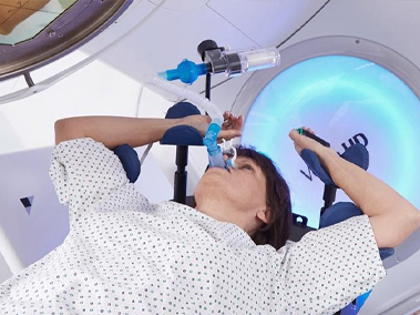
Stereotactic Radiation Therapy For Brain Metastases
Case 1 -Single Brain Metastases
A 55 Years old lady was diagnosed with left breast cancer in August 2015. She was treated with breast surgery, chemotherapy, radiation therapy and hormonal therapy.
She remained disease free for 3 years. In October 2018, she had an episode of seizure. Contrast MRI Brain showed 1.5×1.4 x1.6 cm enhancing lesion in left posterior frontal region of brain with mild surrounding edema suggestive of solitary brain metastases. (Figure 1) MR Spectroscopy showed intense choline peak with reduced NAA suggestive of mitotic etiology. Patient also underwent FDG whole body PET CT scan which showed that brain was the only site of metastases.
Figure 1- Brain MRI – Contrast T1 Sequence showing 1.5×1.4 x1.6 cm intensely enhancing nodule in left posterior frontal region with mild surrounding edema
Management
Neurosurgical resection of brain lesion was ruled out in view of location in close vicinity of motor and speech area.
She was then treated with stereotactic brain radiation therapy. He head was fixed in a non- invasive frame/mould. Radiation therapy (RT) was delivered in 3 fractions/sittings over 3 days using stereotactic radiosurgery (SRS) technique under image guidance. (Figure 2 and 3)
After brain radiosurgery, her hormonal treatment was changed in form of another pill. She was not given chemotherapy as her previous breast cancer was hormone receptor positive.
Follow up
Post brain SRS, patient has a follow up of close to 2 years and she is disease free. She has no neurological deficit and she continues to be on hormonal treatment.
Case 2 –Multiple Brain Metastases
Mr NK, 54 years old gentleman from Kashmir, diagnosed case of metastatic renal cell carcinoma presented to us at the beginning of Corona pandemic with complaints of weakness of right and upper limb.
Clinically he was well preserved except for grade 3 power in right and upper limbs suggestive of right hemiparesis.
His MRI Brain showed total 10 SOLs in brain suggestive of metastases and largest was 1.2 cm in diameter and was causing hemiparesis. (Figure 4)
Figure 4 – Multiple brain metastases as seen on MRI Brain
We treated all his brain lesions with SRS delivered over 3 days and avoided whole brain radiation therapy. (Figure 5 &6)
Immediate after radiation therapy his power on right side improved to 4/5. Since it was a short treatment done over only 3 days, he could go back to Kashmir safely.
Figure 5 and 6 – SRS beam arrangement and dose distribution
Discussion
Till few years back standard treatment of brain metastases was whole brain radiation therapy (WBRT). A new standard of care for brain metastases is to give focal stereotactic radiation therapy. There are several advantages of SRS over WBRT including short treatment, better local control of metastases, no neurocognition decline and no hair loss.
Radiation Therapy For (Left) Breast Cancer With Cardiac / Heart Protection
Breast Cancer is one of the most common cancers among women. Good news is, that many more patients today are long term breast cancer “survivors” with good quality of life because of advances in treatment.
Standard local treatment for breast cancer includes mastectomy or breast conservation surgery followed by chemotherapy and radiation therapy (RT).
Radiation therapy is an essential part of breast cancer treatment as it has been shown to decrease re – occurrence of cancer in breast/chest as well as improve survival rates of patients.
In radiation therapy for left breast cancer, there has always been big concern about safety of radiation doses to heart.
Fortunately, radiation therapy techniques have improved markedly in last few years with special attention to make it more focused and precise to decease its side effects.
Department of Radiation Oncology at Max Super specialty hospital, Shalimar Bagh, is a comprehensive department equipped with latest high end equipments like TrueBeam STx Linear Accelerator, 4 D CT simulator and HDR Brachytherapy to safely do heart sparing left breast cancer RT.
We are equipped with several techniques to protect heart and Lungs while treating breast cancer patients with radiation therapy some of which are mentioned below.
DIBH is an excellent technique technique for protecting heart and lung while doing radiation therapy for left breast cancer.
Prone Position:
At times patients are treated in prone position such that breast falls away from chest wall and hence increases distance from the heart to chest wall/breast. (Figure 3) This also helps considerably in decreasing doses to heart.
Partial Breast Irradiation (PBI):
In case of early breast cancers with favourable molecular profile, sometimes instead of treating whole breast, only small area around the breast tumour is treated with radiation therapy. This is called as partial breast radiation which also significantly reduces dose to heart.
Breast Cancer Radiation Therapy
Radiation Therapy (RT) for breast cancer has seen a long journey precision and focused therapy. RT is an important part of breast cancer management which improve both local control of cancer as well as patients survival.
In earlier times it was a treatment delivered over span of 5-7 weeks with unavoidable high doses of radiation going to underlying heart and lung. Now in the era of precision/ focused radiation therapy technique like IGRT and IMRT, in majority patient’s radiation is delivered over 3-4 weeks with much more safety for heart and lung.
In recent years precision of radiation therapy has further improved with several techniques to protect heart and lung with the most important being DIBH (Deep Inspiratory Breath Hold). In DIBH radiation is delivered in 4D fashion with respiratory gating to keep heart away while delivering radiation therapy.
Further improvement is that in selected patients whole radiation therapy treatment can be delivered over just 4-5 days (rather than weeks) using a technique called APBI (Accelerated Partial Breast Irradiation).
Breast Cancer APBI using intraoperative brachytherapy
Overall breast cancer radiation therapy has become a much shorter treatment with better safety and precision.
Dr Vineeta Goel is currently Director and Head Radiation Oncology at Cancer Institute, Shalimar Bagh. At Fortis Hospital Shalimar Bagh we routinely practice all these advanced techniques for treatment of cancers.
#BreastCancer #RadiationTherapy #APBI #BreastCancerRadiationTherapy
Stereotactic Radiotherapy
Conventional radiation therapy for cancers often results in some collateral damage to nearby healthy tissues. Stereotactic Radiotherapy (SRT) is defined as a method of external beam radiotherapy that use principles of stereotaxy to accurately deliver high radiation dose to a tumour/ target in one or few treatment fractions (1 to 5) without much radiation dose and side effects to the surrounding healthy tissues.
When this technique is used to treat brain tumours (primary /secondary) then it’s called as Stereotactic radiosurgery/ radiotherapy (SRS/SRT). While when this same technique is used for treating tumours outside brain/extra cranial , it’s called as SBRT (Stereotactic Body Radiation Therapy).
SRT is used to treat both primary cancers (where the cancer started such as the lung or the prostate), or where a cancer may have spread to other body organs (secondary cancers or metastases). It is also addressed as SABR (Stereotactic ablative radiation therapy), as it involves the delivery of much higher and near ablative doses of radiation to cancer.
How is SRT different from other forms of RT?
• SBRT involves high precision, image guided focused dose deliveredto the target with rapid dose fall off– translates in to higher control rates
• SBRT has highly conformal radiation with rapid dose fall off to avoid/minimize radiation to healthy normal tissues – minimal side effects
• Larger doses per fraction (typically ≥ 8 Gy per fraction) to ablate the tumor- helps in overcoming radioresistance if any
• Fewer treatment fractions (typically 1-5 fractions) as compared to typical of 15-35 fractions in conventional RT
• Intra-fraction motion management wherever applicable for hitting at the accurate target every day with RT
What are the indications of using Stereotactic Radiation Therapy?
SRT can be used at various sites including
- Primary Brain Tumours like Meningioma, Schwannomas etc.
- Secondary Brain Tumour – Brain Metastases
- Medically inoperable early-stage primary lung cancers (T1/2N0MO)
- Lung metastasis (up to 5-8 in number)
- Spine metastasis
- Primary (Hepatocellular Carcinoma) and secondary liver tumours
- Inoperable/recurrent carcinoma pancreas
- Early low risk prostate cancer
Stereotactic RT delivers high ablative doses precisely to tumour which results in better tumour control and quick symptomatic relief to patients.
Dr. Vineeta Goel, Director Radiation Oncology at Fortis Hospital Shalimar Bagh is the best doctor for STEREOTACTIC RADIOTHERAPY.
# Stereotactic radiation therapy #SRS #SRT #SBRT # Brain Metastases # Liver tumour # Liver Metastases # Spine metastases # Bone Metastases # Lung metastses
Stage IV Breast Cancer With Paralysis Of Lower Limbs
A 48 years old woman presented to us with paralysis of 4 days duration. She also gave a history of upper backache since past 5 months. She had a past history of being treated for right breast cancer with surgery, chemotherapy and hormonal therapy 7 years back. Clinically both her breasts and lymph node regions were normal. Neurologically her power in both lower limbs was reduced to 1/5 and rest everything was normal. We did a MRI spine for her which showed D4 Vertebral metastases with collapse and cord compression. She also underwent whole body PET CT scan which showed that she had multiple bone metastases and her rest body organs were free of cancer.
Patient was jointly evaluated by team of neurosurgeons, radiation and medical oncologists. She was taken up for neurosurgical tumour decompression and spine stabilization in view of recent onset paralysis and metastases being confined only to bones (which carries better prognosis as compared to patient having metastases in lungs or liver). After surgical decompression, her power in lower limbs improved to 2/5.
Surgery also yielded histopathology and biopsy which confirmed it to be metastases from breast cancer with strong hormonal positivity for Estrogen and Progesterone receptors.
Post- surgery she was started on hormonal tablets for breast cancer. Since her breast cancer was confined only to bones and it had strong positivity for hormonal receptors, she did not require any chemotherapy. She was planned for Stereotactic Body Radiation Therapy (SBRT) to affected D4 vertebrae. Her Radiation Therapy was planned using combined information from CT Myelogram and MRI Spine to spare spinal cord and focus high radiation doses only on cancer affected region. This radiation therapy was given over 5 days. She also underwent regular physiotherapy. As a result of this well planned intensive treatment and rehabilitation, she was up on her feet again.
Total Skin Electron Beam Therapy
Total Skin Electron Beam Therapy (TSET) is a radiation technique which is used to treat patient’s entire/total skin surface using electron beams .
TSET is used to treat Cutaneous T-Cell Lymphoma and/or Mycosis Fungoides. Mycosis Fungoides (MF) is a malignancy of helper T cells and accounts for 50% of all cutaneous lymphomas. Clinically it can present as erythematous and scaling skin patches which are generally annular or arc shaped. These later on progress to form infiltrative scaling plaques, nodules and sometimes have superficial ulcerations.
Aim while treating such type of cutaneous lymphomas is to deliver a therapeutic prescribed dose to entire skin of the body to a shallow depth by sparing the deep seated tissues and organs.
Technically TSET is a complex treatment and requires experienced team of Radiation Oncologists, Medical Physicists and Technologists to carry it out well. TSET requires complex dosimetry and quality assurance processes before and during treatment.
Everyday Patients nail bed and eyes are shielded before treatment using special applicators and lead goggles. (Fig 4)
At Max Hospital, Shalimar Bagh we have been treating patients of mycosis fungoides/cutaneous lymphomas with TSET regularly and with very rewarding results.
For each treatment, patients are asked to stand in different positions, alternating daily (total 6 positions) in front of Linear Accerlator. Each position exposes entire patient’s skin to the radiation beam of low energy electrons. (Figure 2-3)
POSSIBLE SIDE EFFECTS OF TSET AND PREVENTIVE MEASURES:
- Itchy skin – keep skin moisturized, wear cotton cloths
- Dry cracking skin – apply moisturizer
- Sun sensitivity- wear sunglass , cap and full gown
- Fatigue – take rest and eat healthy food
- Temperature regulation- Avoid humid hot weather and drink plenty of water
- Partial temporary hair loss
Treatment Outcomes
TSET is the most effective treatment modality for MF. It is a technically challenging and demanding treatment modality and requires stringent quality assurance and team work.
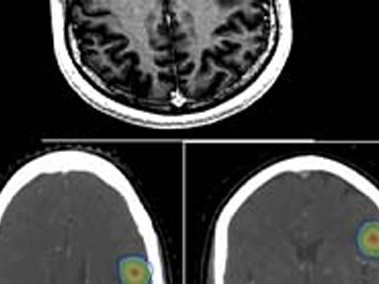
Solitary Brain Metastases From Breast Cancer-Treatment With Stereotactic Radiation Therapy
A 54 Years old lady was diagnosed with left breast cancer in August 2015. Her breast biopsy confirmed it to be Invasive Ductal Carcinoma (IDC) with strong positivity for hormonal receptors (Estrogen and Progesterone) and negativity for Her 2 Neu (Epidermal Growth Factor Receptor). She underwent Left Breast Conservation surgery followed by chemotherapy and Radiation Therapy. After completion of radiation therapy, she was started on oral hormonal therapy treatment – in form of daily tablets.
She remained disease free for 34 months (2 years 10 months), when in October 2018, she had an episode of seizure. Clinically she was well preserved with ECOG performance status of 1, breast cancer was clinically controlled and she was neurologically intact. Contrast MRI Brain showed 1.5×1.4 x1.6 cm intensely enhancing nodule in left posterior frontal region with mild surrounding edema suggestive of solitary brain metastases. MR Spectroscopy showed intense choline peak with reduced NAA suggestive of mitotic etiology. Patient also underwent FDG whole body PET CT scan which showed that brain was the only site of metastases.
Diagnosis
Her final diagnosis was Solitary Brain Metastases from Left Breast Cancer after disease free interval of 34 months in a 54 years old well preserved lady.
Management
Patient was evaluated for surgical resection with the idea that it would also give histo biopsy specimen to repeat hormonal and other IHC markers profile to decide systemic treatment. Her functional MRI brain showed motor activation area in close proximity to metastases and Broca’s area close to FLAIR abnormality. Neurosurgical resection of brain lesion was ruled out in view of location in close vicinity of motor and speech area.
She was then treated with stereotactic brain radiation therapy. He head was fixed in a non- invasive frame/mould. Since her lesion was lying in eloquent area (close to motor and speech cortex), her radiation therapy (RT) was delivered in 3 fractions instead of single fraction RT. She was treated with 27Gy in 3 fractions to brain metastases using stereotactic radiosurgery (SRS) technique under image guidance.
After brain radiosurgery her hormonal treatment was changed in form of another pill. She was not given chemotherapy as her previous breast cancer was hormone receptor positive.
Follow up
Post brain SRS, patient has a follow up of one year 3 months (15 months) and she is disease free. She has no neurological deficit and she continues to be on hormonal treatment. Her repeat MRI Brain showed small residual non enhancing lesion of 6×7 mm and there are no new cranial or extracranial lesions.
Discussion
In past decade, there has been a major shift in approach to management of metastatic cancers with an attempt to potentially cure especially oligo (few/limited) and solitary metastases. This potentially curative treatment approach requires systemic treatment along with aggressive local treatment of metastases either with surgery or ablative high dose radiation therapy with stereotactic approaches (SRS or SBRT).
Till few years back standard treatment of brain metastases was whole brain radiation therapy (WBRT). A new standard of care for brain metastases is to give focal stereotactic radiation therapy. There are several advantages of SRS over WBRT including short treatment, better local control of metastases, no neurocognition decline and no hair loss.
Intra Operative Radiation Therapy (IORT) With Brachytherapy
Intraoperative radiation therapy (IORT) is single dose of focused radiation therapy delivered at the time of surgery.
Brachytherapy is an internal form of radiation therapy and is considered the most conformal (focused/precise) technique.
Brachytherapy when use intraoperatively becomes further focused and spot on.
In cancer treatment, there are several indications of using IORT, with the commonest being
- Recurrent lymph node within the region previously treated with radiation therapy. These lymph nodes can be in neck, axilla (arm pit) or abdomen.
- Recurrent cancer in primary (or site of origin of cancer) where either previously radiation has been done or in sites where it’s difficult to get surgical margins clear at microscopic level (at the level of cells). Common cancers where it is used are rectal cancer, mouth cancers and some pediatric cancers.
Procedure
Pre-operatively patient is jointly evaluated by Surgical and Radiation Oncologist in OPD to take decision regarding need for IORT.
Surgery is done in a special OT with brachytherapy machine in Operation theatre. After surgical removal of tumour, IORT applicator is placed and secured (stitched) to tumour bed (region from where tumour is removed). A quick IORT brachytherapy plan is generated depending on size of applicator and radiation dose to be delivered (generally takes 10 minutes). After planning IORT applicator is connected to brachytherapy machine and radiation therapy is delivered usually over 20-30 minutes. Brachytherapy applicator is then removed and surgical oncologist completes surgery.
Advantages
IORT with Brachytherapy is the most focused and precise form of radiation therapy as its penetration is only about a centimeter deep to where applicator is placed.
It gives high dose to tumour bed with minimum dose to surrounding normal organs.
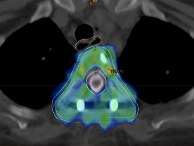
Management Of Intracranial Non Germinomatous Germ Cell Tumour (Choriocarcinoma) Brain With Chemotherapy And Cranio Spinal Radiation Therapy
18 Years old college student presented to hospital with complaints of severe headache, blurring of vision, vomiting and episode of unconsciousness. Clinically patient was conscious, alert but very irritable with normal sensory motor function. He underwent initially CT Head followed by contrast MRI Brain which showed 30x 41 x 39 mm heterogeneous lesion in posterior aspect of third ventricle with contiguous parenchymal infiltration with radiological diagnosis of pineal germ cell tumour.
Subsequently his serum ß HCG was found to be 1, 11, 505 mIU and AFP of 1.36 ng/ml. He underwent CSF sampling which showed no malignant cells on cytology with CSF ß HCG of 1, 40, 614 mIU/ml and AFP of <0.5 ng/ml. His MRI Spine screening was also normal.
Diagnosis
Based on his radiological picture coupled with raised serum and CSF ß HCG with normal AFP, diagnosis of Primary CNS non seminomatous germ cell tumour (NSGCT) with subtype Choriocarcinoma was made.
Management
Patient initially underwent VP shunt control symptoms of raised intracranial pressure and was then started on chemotherapy. After completing four cycles of chemotherapy his serum and CSF ß HCG decreased significantly to 8.4 and 68.6 mIU/ml respectively. His repeat MRI Brain showed much smaller residual disease. Patient also showed much improvement symptomatically.
He was then treated with Cranio Spinal Irradiation (CSI) with Intensity Modulated Image Guided Radiation Therapy (IM IGRT) to a dose of 30.6Gy in 17 fractions followed by local tumour boost to 19.8Gy over 11 fractions. This whole treatment was carried out on TruBeam STX Linear Accelerator at Max Hospital, Shalimar Bagh. Patient tolerated CSI radiation therapy well with grade II haematological and grade I GI toxicity. Patient completed his entire treatment in July 2018.
Follow up
Patient has been on regular follow up for last 18 months and his diseases remains in remission clinically, radiologically and biochemically and he has resumed back his studies in college.
Discussion
Primary Choriocarcinoma of brain is a rare tumour. Biopsy is generally not mandatory for its confirmation as diagnosis is generally made by combination of radiological picture along with raised serum and CSF ß HCG. Surgical resection of tumour is also generally not required as this tumour is very chemo and radio sensitive. Response to treatment is generally done by repeating serum and SCF ßHCG values which should show declining values.
Radiation therapy target for NSGCT Brain includes entire brain and spinal axis including leptomenninges and is termed as cranio spinal radiation. It’s a large and irregular target volume spanning almost across patient’s whole body. Covering cranio spinal axis with radiation therapy also comes with challenge of sparing all organs of body (lung, heart, kidney, esophagus) and cranial structures like eyes, lens, cochlea etc.
With present radiation therapy techniques, it is possible to treat this irregular target volume of brain and spine using combined technique of intensity modulated image guided radiation therapy (IM- IGRT). IMRT helps us to focus radiation doses at correct region while saving adjacent normal organs. IGRT involves using on board Cone beam CT Scan to verify patient positioning every day before radiation therapy.
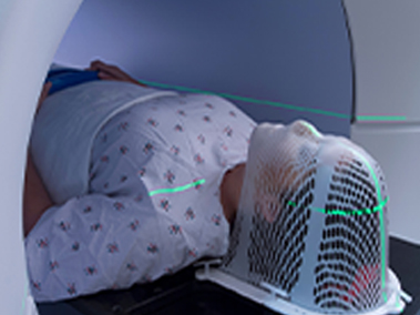
Stereotactic Radiotherapy For Brain Metastases
Spread of cancer to brain called as brain metastases is known to occur in few cancers. All patient with brain metastases require radiation therapy to brain.
Traditional treatment for brain metastases is whole brain radiation therapy over 1-2 weeks. Whole brain radiation therapy is associated with few adverse effects like temporary hair loss, somnolence (tendency to sleep more) and some decline in cognition.
We try to avoid these side effects by using IM-IGRT techniques where we spare some critical areas of brain called hippocampus to avoid cognition decline (called as Hippocampal sparing whole brain RT). In IMRT it is also possible to spare the scalp (skin and hair lining of head) to minimize hair loss.
Good news is that patients with limited /few brain metastases can avoid whole brain RT (and it’s side effects) by focusing radiation therapy only on metastases by a technique called as Stereotactic Radiosurgery (SRS).
In SRS a tight stereotactic mask is made for patient to avoid any movement of head while on treatment. Computerized plan is made using CT scan and MRI together to focus radiation only on metastases. SRS has several advantages like shorter treatment time (2-5 days), minimal hair loss and no decline in cognition.
At Max Institute of Cancer Care, Shalimar Bagh, we are routinely doing SRS and Hippocampal sparing whole brain radiation therapy for suitable patients with brain metastases. Dr Vineeta Goel is an expert radiation oncologist with large experience of SRS and Hippocampal sparing whole brain RT for brain metastases.
Heart Sparing Radiation Therapy
Heart Sparing Radiation Therapy for (Left) Breast Cancer
Breast cancer is one of the most common cancers among women. Good news is, that many more patients today are long term breast cancer “survivors” with good quality of life because of advances in surgery, chemotherapy and radiation therapy.
Standard local treatment for breast cancer includes mastectomy or breast conservation surgery followed by systemic treatment and radiation therapy.
Radiation therapy is a vital part of breast cancer treatment as it has been shown to decrease local In breast cancer radiation therapy, apart from concern about bystander doses to underlying lungs there has always been big concern about safety of radiation doses to heart especially for left breast cancer patients.Fortunately, radiation therapy techniques have improved markedly in last few years with special attention to make it more focused and precise to decease its side effects. Department of Radiation Oncology at Max Super specialty hospital, Shalimar Bagh, is a comprehensive department equipped with high end equipment like TrueBeam STx Linear Accelerator, 4 D CT simulator and HDR Brachytherapy. (Fig 1)
We are equipped with several techniques to protect heart and Lungs while treating breast cancer patients with radiation therapy some of which are mentioned below.
Deep Inspiratory Breath Hold (DIBH):
Deep inspiration breath hold (DIBH), is a radiation therapy technique where patient is trained to hold breath in deep inspiration. By holding breath in deep inspiration, patients fill extra air in lungs which moves heart away from your breast/ chest and decreases dose to heart and lungs (as shown in figure 2). Radiation beams are synchronised with patient’s breathing cycles, such that radiation is delivered only when patient is in DIBH using RPM (Respiratory Motion Management Camera). This is also called as 4 D Radiation Therapy where 4th dimension is time.
Prone Position:
At times patients are treated in prone position such that breast falls away from chest wall and hence increases distance from the heart to chest wall/breast. (Figure 3) This also helps considerably in decreasing doses to heart.
Partial Breast Irradiation (PBI):
In case of early breast cancers with favourable molecular profile, sometimes instead of treating whole breast, only region around the breast tumour is treated with radiation therapy. This is called as partial breast radiation which also significantly reduces dose to heart. (Fig 4)
Conclusion:
With the advent of present technologies, radiation therapy has become as safe for left breast cancer as its for right side. Dr Vineeta Goel is the best radiation oncologist for heart sparing radiotherapy for breast cancer. She is currently Director and Head Department of Radiation Oncology at Fortis Hospital, Shalimar Bagh, Delhi. She has over 20 years of experience in the field of oncology and radiation oncology. Prior to joining Fortis Healthcare, Dr Vineeta was working as Associate Director Radiation Oncology at Max Healthcare for last 12 years. She has been trained from prestigious Tata Memorial Hospital, Mumbai and has also worked there as Assistant Professor. She describes herself as “passionate and compassionate” oncologist.
Department of Radiation Oncology at Fortis Hospital, Shalimar Bagh is a comprehensive department equipped with Elekta Versa HD, CT simulator and HDR brachytherapy.
Carcinoma Cervix With Intrauterine Fibroids
56 years old lady with no comorbidities was investigated for postmenopausal bleeding PV. Clinically she had growth involving cervix with bulky uterus and bilateral parametrium medially infiltrated by disease. Biopsy from cervical mass showed moderately differentiated squamous cell carcinoma. Contrast MRI abdomen showed bulky cervical mass with multiple subserosal, submucosal and intramural fibroids with largest fibroid measuring 8.6 x 7cm. There was no lymphadenopathy; both liver and kidneys were normal. Her Chest X-ray was unremarkable.
Diagnosis
Her final diagnosis was squamous cell carcinoma cervix FIGO stage II B with multiple large intra uterine fibroids.
Management
Standard of care for this stage cervical carcinoma is concurrent chemotherapy and external beam radiation therapy for 5 weeks followed by intracavitary brachytherapy. The challenge before us in this was the large intra uterine fibroids would prevent successful placement of intracavitary brachytherapy applicator. Surgery was already ruled out due to bilateral medial parametrium infiltration by disease.
After discussion in multispeciality tumour board we decided to treat this patient with 5 weeks of concurrent chemoradiotherapy (CTRT). She received weekly Injection Cisplatin 35 mg/m2 along with 45 Gy/25 Fractions of radiation therapy to cervix, uterus and pelvic lymph nodes with image guided Intensity Modulated Radiation Therapy (IG- IMRT).
Patient tolerated CTRT well and clinically had >80% regression in cervical mass. Repeat MRI at completion of CTRT also showed >80 disease regression with small residual diseases with bilateral medial parametrium infiltration.
The patient was next taken up for Interstitial Brachytherapy along with intra uterine tube placement under USG guidance. USG helped us navigate our needles and tubes in correct position despite multiple large obstructing intrauterine fibroids. This whole procedure was done under spinal anesthesia with epidural catheter for maintain analgesia. Patient with stood this procedure well.
Follow Up
Post treatment she has a follow up of one year and she is disease free.
Discussion
Radiation Therapy has been the treatment modality of choice for management of cancer cervix for more than a century. In last two decades radiation therapy with weekly concurrent chemotherapy (CTRT) has become the new standard of care.
Success of radiation therapy in providing high cure rates in carcinoma cervix is essentially due to use of brachytherapy. Brachytherapy allows us to give high doses of radiation inside tumour region with very less radiation dose to surrounding normal organs. Treatment of carcinoma cervix remains incomplete without brachytherapy. Presence of intrauterine fibroids is considered a relative contraindication for treating patients with radiation therapy as brachytherapy is generally not feasible. In this patient, we could manage successful brachytherapy as we used intraoperative USG guidance and we replaced traditional intracavitary brachytherapy with interstitial procedure. Interstitial brachytherapy gives us more freedom using needles to adequately cover the entire tumour with adequate radiation doses.
Stage IV Breast Cancer With Paralysis Of Lower Limbs
A 48 years old woman presented to us with paralysis of 4 days duration. She also gave a history of upper backache since past 5 months. She had a past history of being treated for right breast cancer with surgery, chemotherapy and hormonal therapy 7 years back. Clinically both her breasts and lymph node regions were normal. Neurologically her power in both lower limbs was reduced to 1/5 and rest everything was normal. We did a MRI spine for her which showed D4 Vertebral metastases with collapse and cord compression. She also underwent whole body PET CT scan which showed that she had multiple bone metastases and her rest body organs were free of cancer.
Patient was jointly evaluated by team of neurosurgeons, radiation and medical oncologists. She was taken up for neurosurgical tumour decompression and spine stabilization in view of recent onset paralysis and metastases being confined only to bones (which carries better prognosis as compared to patient having metastases in lungs or liver). After surgical decompression, her power in lower limbs improved to 2/5.
Surgery also yielded histopathology and biopsy which confirmed it to be metastases from breast cancer with strong hormonal positivity for Estrogen and Progesterone receptors.
Post- surgery she was started on hormonal tablets for breast cancer. Since her breast cancer was confined only to bones and it had strong positivity for hormonal receptors, she did not require any chemotherapy. She was planned for Stereotactic Body Radiation Therapy (SBRT) to affected D4 vertebrae. Her Radiation Therapy was planned using combined information from CT Myelogram and MRI Spine to spare spinal cord and focus high radiation doses only on cancer affected region. This radiation therapy was given over 5 days. She also underwent regular physiotherapy. As a result of this well planned intensive treatment and rehabilitation, she was up on her feet again.
This case exemplifies importance of multi disciplinary treatment, tumour board, newer radiation techniques like SBRT and quality of life of patients.
Radiation Therapy In Cancer Management
What is Radiation Therapy and what does it do?
Radiation Therapy is use of high energy Ionization radiation (often X Rays ) to kill a cancer cell by damaging its DNA. Goal of Radiation Therapy (RT) is to use focused beams of radiation to kill cancer cell with as little risk as possible to normal cells. Radiation treatment, like surgery, is a local treatment. It affects the cancer cells only in a specific area of the body.
Timing of RT
RT can be used before surgery to shrink a tumor, called as neoadjuvant/pre operative RT (Pre op RT). It may be used after surgery to stop growth of cancer cells that may remain called as adjuvant RT (Post op RT). At times radiation is used alone with curative intent which is called as Radical RT. It can also be used at the time of surgery known as Intraoperative Radiation (IORT).
Types of RT
When the source of radiation is from a machine outside the body, it’s called as External Beam Radiation therapy (EBRT). When radioactive material is placed close to tumour or area harboring cancer cells it’s called as Brachytherapy.
What is EBRT?
EBRT is usually given during outpatient visits (OPD Treatment) to a hospital. In this, a machine (also called as Linear Accelerator/LA) directs the high energy rays at the tumor bearing area within body. Linear Accelerators have the capability of delivering RT by various techniques like IMRT (Intensity Modulated Radiation Therapy), IGRT (Image Guided Radiation Therapy), SRS (Stereotactic Radiation Therapy) and SBRT (Stereotactic Body Radiation Therapy).
What is IMRT?
Intensity-modulated radiation therapy (IMRT) refers to a technique of focusing radiation therapy at cancer bearing area using computer based optimization process to carefully create a gradient or a dose fall-off between the cancer/target tissues and the surrounding normal tissues. This fall of dose saves the surrounding normal organs effectively. Therefore, these techniques offer the prospect of increasing the cancer control probability while decreasing the side effects.
What is IGRT?
Image guided radiation therapy (IGRT) is classically defined as radiation therapy that is delivered only after verification of position of structures of interest by performing either an X-Ray or CT based image. The same are done by an imaging system mounted on the linear accelerator itself. IGRT technically includes IMRT and hence also called as IM- IGRT (Intensity-modulated Image guided radiation therapy)
What is SBRT?
Stereotactic body radiation therapy (SBRT) refers to use of principles of stereotaxy to identify and pin pointedly deliver precisely deliver intense doses of RT to only to cancer/tumour area. SBRT is essentially similar to Stereotactic Radiosurgery (SRS) brain except that term SBRT used when we are targeting areas outside brain. Since SBRT involves delivery of higher intensity of radiation doses in shorter time, it also known as SABR (Stereotactic Ablative RT). SBRT is generally done by combining IMRT and IGRT together. SBRT generally has fewer sitting /fractions of radiation as compared to conventional IMRT or IGRT.
Your Radiation Oncologist will discuss all these techniques and how they are relevant for your cancer and its treatment.
What is more important for successful radiation therapy- Machine or team behind machine?
Both machines and team behind machines are equally critical in successful delivery of RT.
While technology always opens the door, it is always a skillful doctors who can get inside it.
Post mastectomy pain—Less discussed but common problem
Mastectomy can be associated with some annoying symptoms like
- Tightness around the chest
- Chronic chest pain
- Stiffness and reduced shoulder movement
- Shoulder imbalance and associated upper back pain
Radiation therapy after mastectomy can aggravate all these symptoms. Some of these symptoms can have an impact on quality of life.
However, both these treatments are lifesaving essential treatments.
Why do these symptoms happen after mastectomy?
- During mastectomy along with breast, some chest muscles are removed because of which strength and flexibility of chest reduces.
- Arm pit fat pad is removed, and some nerves are cut during lymph node removal surgery which also leads to pain and reduced shoulder movement.
- Lymph is a white fluid which flows through out our body. After breast and arm pit surgery, its free flow/circulation gets impacted leading to collection of fluid around chest and arm called as lymphedema which can also causes pain and heaviness of chest and arm.
- After axillary/arm pit surgery, some women can develop, rope or cord like structure just under the skin in the area under your arm called as axillary web syndrome (AWS).
- Radiation therapy after mastectomy leads to fibrosis or in simpler terms shrinkage of fat and muscles left after mastectomy which makes symptoms of tightness, chest pain worse.
What can we do to reduce intensity of these symptoms and prevent them from getting bad to worse? Few suggestions are
- Regular and graded physical activity/exercise of affected shoulder, upper back, arm and neck to strengthen your remaining muscles and improve flexibilty. Regular physical therapy also helps prevent lymphedema/ swelling in arm. It should be learned with the help of a professional physiotherapist.
- Regular massage and light rubbing of chest and arm pit scar using some thick cream in upwards, circular and lateral scar movement.
- Use of adequate size and weight prosthesis to maintain balance and posture of shoulder and back.
What to do if symptoms do not get better after above remedies also?
- Consult your oncologist and discuss with him/her.
- Your oncologist might add some suitable pain killer medication for you.
- Sometimes even acupressure is known to help in reducing pain.
- If pain is due to lymphedema, then your oncologist might refer you to a physical therapist to learn manual drainage of accumulated lymph
Prevention of all these symptoms which occur due to “after effect” of cancer treatment is the key.
Author
Dr Vineeta Goel
Director and Head
Department of Radiation Therapy
Fortis Hospital
Shalimar Bagh, Delhi
# Breast Cancer
# Mastectomy
# Pain and swelling after breast cancer surgery
Left Breast Cancer Radiation Therapy Respiratory Gating To Save Heart
Safety of modern breast RT for lungs and heart
- Modern Radiation therapy with IMRT/IGRT is much safer
- Small risk of cardiac ds (5-6% risk at 20 years) especially for left breast cancers
- Pneumonitis <2%
Are there radiation techniques for better heart sparing?
- Partial Breast Radiation
- Prone Treatment
- Use of respiratory gating- 4D RT
Gated Breast Radiation Therapy- 4D RT
- Gating refers to 4D RT- synchronizes RT with breathing cycle
- Pts are coached to take deep breath and then hold that level of inspiration during every radiation treatment
- BH can increase distance between heart and target volume
- Beam on only during breath hold in deep Inspiration
- Its associated with lower radiation exposure to heart without compromising coverage of disease
Breast Cancer Radiation Therapy- Journey From 5 Weeks to 5 Days Treatment
Radiation Therapy (RT) for breast cancer has seen a long journey precision and focused therapy. RT is an important part of breast cancer management which improve both local control of cancer as well as patient’s survival.
In earlier times it was a treatment delivered over span of 5-7 weeks with unavoidable high doses of radiation going to underlying heart and lung.
Now in the era of precision/ focused radiation therapy technique like IGRT and IMRT, in majority patient’s radiation is delivered over 3-4 weeks with much more safety for heart and lung.
In recent years precision of radiation therapy has further improved with several techniques to protect heart and lung with the most important being ABC (Active Breath Coordinator).
In ABC, radiation is delivered in 4D fashion with respiratory gating to keep heart away while delivering radiation therapy.
Further improvement is that in selected patients whole radiation therapy treatment can be delivered over just 4-5 days (rather than weeks) using a technique called APBI (Accelerated Partial Breast Irradiation).
Overall breast cancer radiation therapy has become a much shorter treatment with better safety and precision.
We routinely practice all these advance techniques at department of radiation oncology, Fortis hospital, Shalimar Bagh.
#Breast Cancer #Radiation Therapy # APBI # Breast cancer Radiation Therapy #DIBH
Intraoperative Radiotherapy
Intraoperative radiation therapy (IORT) is a form of precise and impactful radiation therapy. IORT refers to a single shot of high dose radiation therapy given to the tumour bed (area of cancer) during cancer surgery.
- There are several case scenarios where IORT finds it’s great advantage.
- There is only one way to find its appropriate use and that is discussion within team of oncologists.
- IORT improves the chances of local control of cancer many fold with least side effects.
Sharing two cases where we used HDR brachytherapy based IORT.
Case 1 – Recurrent and previously radiated cancer rectum.
50 years old gentleman with a history of renal transplant and multiple co morbidities developed low rectal adenocarcinoma which was initially treated with pre-operative chemo radiation therapy (CTRT). The patient was planned for surgery post chemo-radiation, however he defaulted the same. One year later the patient presented with local recurrence in rectum infiltrating in adjacent prostate gland. He was treated with neo adjuvant chemotherapy and low dose CTRT with partial response. He then underwent surgery (Abdomino Perineal resection) by a team of surgical oncologist with IORT to tumour bed at prostate. IORT was done using HDR Brachytherapy to the dose of 10Gy single fraction to prostate bed. After 18 months of treatment patient is free of disease and has no major adverse effects.
Case 2 – Locally Extensive Carcinoma Buccal Mucosa
52 years old gentleman presented with buccal alveolar cancer with extension to infra temporal fossa. He was initially treated with neoadjuvant chemotherapy (NACT) with partial response. Intraoperatively disease was infiltrating into pterygoid plates. After tumour resection by surgical oncology team, we did IORT using HDR brachytherapy to a dose of 12Gy in single fraction. After 4 weeks from surgery, patient underwent additional post-operative radiation therapy 60Gy/30 fractions with IM IGRT technique. After 18 months of treatment patient is free of disease and has no major adverse effects.
Interstitial / Intracavitary Brachytherapy For Cervical Cancer
Brachytherapy is a boon of medical science for patients having cervical cancer.
And it needs deep knowledge and extensive equipment for this treatment to be successful.
During intracavitary/Interstitial brachytherapy for cervical cancer treatment, an applicator with few tubes is placed within patient’s tumour and is later loaded with radiation source to give high focused radiation dose.
Brachytherapy for cervical cancer treatment has been a boon for the patients because of the following reasons:
- This technique uses high radiation only on the tumor and only small radiation is applied to the rest of the body.
- Without brachytherapy, treatment of cervical cancer remains incomplete.
- It does not have as much side effect as other treatment methods of cancer, as only the part where the tumor exits are kept under high radiation.
So, these are some of the salient features of this gifted technique of medical science for the treatment of cervical cancer.
This treatment requires experience, knowledge, and skill, so it is needed to be performed by an expert. Dr Vineeta Goel is an expert with vast experience in both Intracavitary and Interstitial Brachytherapy for cervical cancer treatment in Delhi. Dr. Vineeta Goel is presently Director & Head Department of Radiation Oncology at Fortis Hospital, Shalimar Bagh, Delhi.
She is regarded as one of the best oncologist because of her vast knowledge and experience in this difficult field of medical science. She has been practicing oncology for the last 20 years. The most important point which testifies her journey of success in this field is the fact that Dr. Vineeta not only cures the patients of the dangerous disease of cancer but also makes them get back to their normal lives. She makes patients mentally prepared and positive, due to which this revolutionary therapy works at an even faster rate. Along with this, she gives preference to quality treatment and satisfaction of her patients.